













- 1st October 2026
Table of Contents
- What Does ALT Actually Tell Us?
- ALT and Liver Fibrosis Are Not the Same Thing
- Can You Really Have Fatty Liver With Normal ALT?
- Does Normal ALT Mean the Fatty Liver Is Mild?
- Why Can ALT Remain Normal Despite Liver Disease?
- What Is MASLD?
- Simple Steatosis, MASH, Fibrosis and Cirrhosis Are Different
- Fibrosis Is One of the Most Important Things to Assess
- Ultrasound Fat Grade and Fibrosis Stage Answer Different Questions
- What Is a “Normal” ALT Anyway?
- ALT Can Also Fluctuate
- What About AST?
- What Is FIB-4?
- A Normal ALT Does Not Make FIB-4 Unnecessary
- How Is FIB-4 Generally Interpreted?
- Age Changes How FIB-4 Should Be Interpreted
- Type 2 Diabetes Changes the Level of Concern
- Obesity Also Matters
- Can Lean People Develop Fatty Liver?
- Prediabetes and Insulin Resistance Matter Too
- High Triglycerides Can Be Another Metabolic Clue
- What About Blood Pressure?
- Sleep Apnoea Can Also Matter
- Which Patient With Normal ALT Deserves More Careful Fibrosis Assessment?
- What Happens if FIB-4 Is Not Clearly Low Risk?
- What Does FibroScan Measure?
- Does Everyone With Fatty Liver Need FibroScan?
- Why Not Perform FibroScan on Everyone?
- Can Someone Have Advanced Fibrosis and Still Feel Completely Well?
- Can Bilirubin Remain Normal Too?
- The Liver Has Considerable Functional Reserve
- How Can Significant Fibrosis Develop When ALT Is Normal?
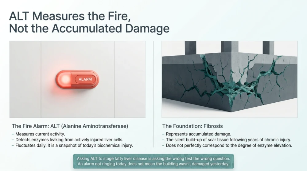
- Think of ALT as Activity and Fibrosis as Accumulated Damage
- Why Fibrosis Matters More Than the Ultrasound Grade
- Can Severe Fatty Liver Exist Without Advanced Fibrosis?
- Fibrosis Is a Major Prognostic Marker in MASLD
- This Is Why the Assessment Usually Happens in Steps
- Understanding FIB-4 Properly
- What Does a FIB-4 Below 1.3 Mean?
- What Does FIB-4 of 1.3 or Higher Mean?
- Why FIB-4 Can Be Misleading in Younger Adults
- Why FIB-4 Can Run Higher in Older Adults
- Do Not Calculate FIB-4 During Acute Illness and Treat It as Chronic Fibrosis Staging
- Can Platelet Count Provide a Clue?
- What Is FibroScan?
- What Does Liver Stiffness Measurement Tell Us?
- What Can Temporarily Increase Liver Stiffness?
- What FibroScan Numbers Are Used in MASLD?
- What Is CAP?
- Can CAP Tell Me Whether I Have MASH?
- Can CAP Tell Me Whether My Liver Is Scarred?
- Why Fasting Before FibroScan Matters
- What Is the ELF Test?
- What About MR Elastography?
- What Is MRI-PDFF?
- Does Anyone Still Need a Liver Biopsy?
- Why Is Liver Biopsy Not Used for Everyone?
- Type 2 Diabetes Deserves Special Attention
- Why Does Diabetes Increase Liver Risk?
- Should Every Person With Diabetes Be Checked for Fibrosis?
- What About Prediabetes?
- Can Significant Fibrosis Occur in Someone Without Diabetes?
- Alcohol Still Matters in Someone With Metabolic Fatty Liver
- Do Not Forget Other Causes of Liver Disease
- Can Weight Loss Improve Fatty Liver?
- Resistance Training or Cardio for Fatty Liver?
- Muscle Matters in Fatty Liver Disease
- What Should Be Monitored Besides ALT?
- Why Cardiovascular Risk Cannot Be Ignored
- What if ALT Becomes Normal After Weight Loss?
- Can Fibrosis Improve?
- Can Cirrhosis Be Present With Normal ALT?
- What Other Clues May Suggest Advanced Liver Disease?
- How Should Fatty Liver With Normal ALT Be Managed?
- Start by Establishing the Fibrosis Risk
- Do Not Treat the ALT Number—Treat the Patient
- Weight Reduction Can Produce Major Liver Benefits
- How Much Weight Loss Should You Aim For?
- For an 80 kg Person, What Do These Percentages Mean?
- But Weight Loss Should Not Mean Muscle Loss
- What Should You Eat for Fatty Liver?
- A Mediterranean-Style Pattern Can Be Adapted to Indian Food
- What Can an Indian Fatty-Liver Plate Look Like?
- Do You Need to Stop Eating Rice and Roti?
- Reduce Added Sugar and Sugar-Sweetened Beverages
- Do You Need a Zero-Sugar Diet?
- Protein Deserves Attention
- Exercise Can Help Even Before Major Weight Loss Occurs
- How Much Exercise?
- Walking After Meals Is Particularly Practical
- Resistance Training Should Not Be Forgotten
- What About Alcohol?
- What About Coffee?
- Do Liver Detox Drinks Help?
- Be Careful With Herbal and “Natural” Liver Supplements
- What About Vitamin E?
- Diabetes Treatment Is Part of Liver Treatment
- What About GLP-1-Based Treatment?
- There Are Now Disease-Directed Treatments for Selected MASH Patients
- Statins and Fatty Liver: A Common Misunderstanding
- How Often Should FIB-4 Be Repeated?
- Does FibroScan Need to Be Repeated Every Year?
- A Practical Monitoring Framework
- When Should You See a Liver Specialist?
- Red Flags That Need Prompt Medical Assessment
- Common Myths About Fatty Liver With Normal ALT
- Myth 1: Normal ALT Means My Liver Is Healthy
- Myth 2: Grade 1 Fatty Liver Means Stage 1 Fibrosis
- Myth 3: If AST and ALT Are Normal, I Do Not Need Fibrosis Assessment
- Myth 4: FibroScan and Ultrasound Are the Same Test
- Myth 5: A High CAP Score Means Advanced Fibrosis
- Myth 6: FIB-4 Above 1.3 Means I Have Advanced Fibrosis
- Myth 7: Only People With Obesity Develop Fatty Liver
- Myth 8: Liver Detox Drinks Can Remove Fat From the Liver
- Myth 9: Fatty Liver Means I Must Stop Eating All Carbohydrates
- Myth 10: Nothing Can Be Done Once Fibrosis Starts
- What I Tell Patients With Fatty Liver and Normal ALT
- A Practical Fatty-Liver Checklist
- Key Takeaways
- References
Fatty Liver With Normal ALT: Can Significant Liver Disease Still Be Present?
Your ultrasound says fatty liver.
You look at your blood tests.
ALT is normal.
AST is normal.
Bilirubin is normal.
So naturally, you may think:
“My liver tests are normal, so the fatty liver cannot be serious.”
This is one of the most important misconceptions I encounter when discussing fatty liver disease.
A normal ALT is certainly preferable to a markedly elevated ALT.
But normal ALT does not prove that the liver is free from significant disease.
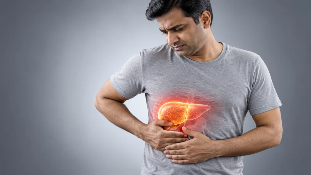
A person can have fatty liver with normal ALT and still have clinically important metabolic dysfunction.
More importantly, some people can have liver fibrosis despite aminotransferase levels that remain within the laboratory reference range.
This is why modern assessment of metabolic dysfunction-associated steatotic liver disease, or MASLD, does not stop after looking at ALT and AST.
The more important question is:
“What is this person's risk of liver fibrosis?”
What Does ALT Actually Tell Us?
ALT stands for alanine aminotransferase.
It is an enzyme found predominantly within liver cells, although it is not exclusively confined to the liver.
When hepatocytes are injured, ALT can leak into the bloodstream.
This is why ALT is commonly included in liver-function panels.
But ALT Is Primarily a Marker of Injury, Not a Direct Measure of Fibrosis
This distinction is fundamental.
ALT can provide evidence that liver cells are being injured.
But it does not directly measure:
- How much fat is present in the liver.
- Whether inflammation is present histologically.
- How much scar tissue has accumulated.
- Whether significant fibrosis is present.
- Whether portal hypertension has developed.
Therefore, asking ALT to tell us the stage of fatty liver disease is asking the wrong test to answer the wrong question.
ALT and Liver Fibrosis Are Not the Same Thing
Fibrosis means accumulation of scar tissue within the liver following chronic injury and repair.
Fibrosis develops gradually.
It is commonly described in stages ranging from minimal or absent fibrosis through progressively more advanced fibrosis and eventually cirrhosis.
ALT does not measure this scar tissue.
A Useful Way to Think About It
ALT asks: Is there biochemical evidence suggesting liver-cell injury?
Fibrosis assessment asks: Has chronic liver disease produced clinically important scarring?
These questions overlap, but they are not interchangeable.
Can You Really Have Fatty Liver With Normal ALT?
Yes.
In fact, this is not unusual.
Many people with MASLD are identified incidentally.
They may undergo an abdominal ultrasound for abdominal discomfort, kidney stones, gallbladder symptoms or a routine health assessment.
The report unexpectedly mentions:
“Fatty changes in liver.”
But ALT and AST may remain within the laboratory reference range.
Normal aminotransferases therefore do not exclude MASLD.
Does Normal ALT Mean the Fatty Liver Is Mild?
Not necessarily.
This is where interpretation becomes more important.
A patient may have:
- Normal ALT.
- Normal AST.
- Normal bilirubin.
- No liver-related symptoms.
and still require assessment for fibrosis depending on their metabolic risk profile.
Normal Blood Tests Can Create False Reassurance
Imagine a 48-year-old patient with:
- Type 2 diabetes.
- Central obesity.
- High triglycerides.
- Hypertension.
- Ultrasound-confirmed fatty liver.
- ALT reported within the laboratory range.
It would be inappropriate to conclude that the liver disease is automatically harmless simply because ALT is normal.
The presence of diabetes and multiple cardiometabolic risk factors makes fibrosis assessment more relevant.
Why Can ALT Remain Normal Despite Liver Disease?
ALT concentration in the bloodstream is influenced by many factors.
It is not a continuously rising gauge that increases in exact proportion to liver damage.
Chronic liver disease is biologically more complicated.
The degree of biochemical enzyme elevation does not perfectly correspond to the amount of fibrosis.
Liver Disease Can Be Quiet
MASLD commonly progresses without obvious symptoms.
Even clinically important fibrosis may remain silent for years.
A person can feel well, work normally, exercise and have no liver pain while chronic liver disease progresses.
This is why symptom-based screening is inadequate.
What Is MASLD?
The terminology surrounding fatty liver disease has changed.
MASLD stands for metabolic dysfunction-associated steatotic liver disease.
It broadly describes hepatic steatosis occurring in association with cardiometabolic risk factors when the condition fits the current diagnostic framework.
The older term NAFLD—non-alcoholic fatty liver disease—is still encountered frequently in previous reports, research papers and patient records.
MASLD Is More Than Fat Stored in the Liver
MASLD frequently occurs alongside:
- Abdominal obesity.
- Insulin resistance.
- Prediabetes.
- Type 2 diabetes.
- High triglycerides.
- Low HDL cholesterol.
- Hypertension.
- Obstructive sleep apnoea.
This is why I prefer to view fatty liver as part of the patient's broader metabolic health rather than as an isolated ultrasound finding.
Simple Steatosis, MASH, Fibrosis and Cirrhosis Are Different
Fatty liver disease exists across a spectrum.
| Stage or Feature | What It Broadly Means |
|---|---|
| Steatosis | Excess fat has accumulated within the liver |
| MASH | Steatotic liver disease with characteristic inflammatory and hepatocellular injury features |
| Fibrosis | Scar tissue has accumulated within the liver |
| Advanced fibrosis | More extensive scarring associated with substantially greater liver-related risk |
| Cirrhosis | Advanced architectural distortion and fibrosis with risk of portal hypertension, liver failure and liver cancer |
Not every person with fatty liver progresses through all of these stages.
Many will never develop advanced liver disease.
The challenge is identifying the smaller group that is at greater risk.
Fibrosis Is One of the Most Important Things to Assess
When discussing prognosis in MASLD, the amount of fibrosis is extremely important.
Greater fibrosis burden is associated with higher risk of liver-related complications and mortality.
This is why current approaches emphasise fibrosis risk stratification rather than simply asking whether ultrasound says “grade 1” or whether ALT is elevated.
“Grade 1 Fatty Liver” Does Not Mean “Stage 1 Liver Disease”
This is another common misunderstanding.
Ultrasound descriptions such as mild, moderate or severe fatty liver refer primarily to the sonographic appearance of steatosis.
They do not reliably tell us the stage of fibrosis.
A report saying “Grade 1 fatty liver” therefore should not automatically be interpreted as “very early liver disease with no scarring.”
Ultrasound Fat Grade and Fibrosis Stage Answer Different Questions
| Test/Measurement | Main Question | Major Limitation |
|---|---|---|
| ALT/AST | Is biochemical hepatocellular injury evident? | Cannot reliably stage fibrosis |
| Conventional ultrasound | Does the liver appear steatotic? | Limited for quantifying fibrosis and can miss lesser degrees of steatosis |
| FIB-4 | Is advanced fibrosis unlikely or does the patient need further assessment? | Risk-stratification tool rather than direct fibrosis measurement |
| Transient elastography | What is the liver stiffness and therefore estimated fibrosis risk? | Results can be influenced by technical and clinical factors |
| Liver biopsy | What does the liver tissue show histologically? | Invasive, has sampling limitations and is not required for most patients |
What Is a “Normal” ALT Anyway?
This question is more complicated than it first appears.
Laboratory reference ranges are not identical.
A value considered within the reference interval at one laboratory may sit above a healthier population-based threshold used in another clinical context.
Reference intervals can also differ according to sex and methodology.
Laboratory Normal Does Not Always Mean Optimal Liver Health
A patient may say:
“My ALT is 38 and the laboratory says normal up to 45, so my liver must be completely healthy.”
That conclusion goes beyond what the test can establish.
ALT should be interpreted with the clinical context, metabolic risk and other liver assessments rather than simply comparing one number with the upper boundary printed on a report.
ALT Can Also Fluctuate
ALT is not necessarily constant.
Values may vary over time.
A patient may have intermittent elevations followed by normal measurements.
Therefore, a single normal result should not automatically erase a previous pattern of abnormal liver biochemistry.
What About AST?
AST stands for aspartate aminotransferase.
Unlike ALT, AST is found substantially outside the liver as well, including in skeletal muscle.
AST therefore also needs context.
Heavy exercise, muscle injury and other conditions can influence AST.
Normal AST Does Not Exclude Fibrosis Either
Just like ALT, AST alone cannot reliably stage MASLD.
However, AST becomes useful when combined with age, ALT and platelet count in a validated fibrosis risk score called FIB-4.
What Is FIB-4?
FIB-4 is a simple non-invasive fibrosis risk assessment calculated from:
- Age.
- AST.
- ALT.
- Platelet count.
It is one of the most widely used first-line tools for assessing the likelihood of advanced fibrosis in people with MASLD.
Why Is FIB-4 Useful?
Most people with fatty liver do not need a liver biopsy.
What clinicians need is an inexpensive first step that helps identify people who are unlikely to have advanced fibrosis and those who require further assessment.
FIB-4 is useful for this purpose.
A Normal ALT Does Not Make FIB-4 Unnecessary
This is particularly important for the topic of this article.
If someone has fatty liver and relevant metabolic risk, fibrosis assessment should not automatically be abandoned because ALT happens to fall within the laboratory reference range.
Risk stratification depends on the overall clinical situation.
How Is FIB-4 Generally Interpreted?
For many adults with MASLD in the usual primary-care age range, a FIB-4 below approximately 1.3 is commonly used to identify a lower probability of advanced fibrosis.
A value at or above this threshold generally leads to secondary fibrosis assessment rather than automatically diagnosing advanced fibrosis.
FIB-4 Is a Triage Tool, Not a Diagnosis
A FIB-4 of 1.4 does not mean:
“You definitely have liver fibrosis.”
It means that the first-line assessment has not confidently placed the patient in the low-risk group and additional evaluation may be appropriate.
Age Changes How FIB-4 Should Be Interpreted
FIB-4 has important age-related limitations.
It performs less reliably in younger adults, particularly below approximately 35 years of age.
In adults over 65, age itself can push the score upward, so an age-adjusted approach is used.
This is why entering numbers into an online calculator without understanding the patient can lead to inappropriate reassurance or unnecessary anxiety.
Type 2 Diabetes Changes the Level of Concern
Type 2 diabetes is one of the most important risk factors for progressive MASLD and advanced fibrosis.
Therefore, a patient with fatty liver and diabetes deserves particular attention even when ALT is normal.
This Patient Should Not Be Reassured by ALT Alone
Consider someone with:
- Type 2 diabetes for 10 years.
- Abdominal obesity.
- Hypertension.
- High triglycerides.
- Ultrasound-confirmed fatty liver.
- ALT of 28 U/L.
The correct conclusion is not:
“ALT is 28, therefore the liver is fine.”
The more appropriate question is:
“Has this patient been appropriately assessed for fibrosis risk?”
Obesity Also Matters
Increasing adiposity, particularly visceral adiposity, is closely associated with MASLD.
But BMI alone does not tell the entire story.
Waist circumference and the presence of other metabolic complications add useful information.
Fat Distribution Matters
Two people with the same BMI can have different amounts of visceral fat, liver fat and skeletal muscle.
Their metabolic and liver risk may therefore be different.
Can Lean People Develop Fatty Liver?
Yes.
MASLD is not restricted to people with obvious obesity.
Some individuals with a BMI within the conventional normal range develop hepatic steatosis and metabolic dysfunction.
This is particularly relevant in South Asian populations, where cardiometabolic abnormalities can occur at comparatively lower BMI levels.
Do Not Dismiss Fatty Liver Because the Patient “Doesn't Look Overweight”
Waist circumference, visceral adiposity, glucose regulation, triglycerides, blood pressure, family history and physical activity all deserve consideration.
Prediabetes and Insulin Resistance Matter Too
MASLD is closely linked with insulin resistance.
When adipose tissue, liver and skeletal muscle become less responsive to insulin, abnormalities in glucose and lipid metabolism can contribute to hepatic fat accumulation.
This is why fatty liver frequently occurs alongside prediabetes and type 2 diabetes.
High Triglycerides Can Be Another Metabolic Clue
Elevated triglycerides commonly accompany insulin-resistant states and MASLD.
A patient with fatty liver, abdominal obesity, high triglycerides and hypertension has a different metabolic risk profile from someone with an isolated incidental imaging finding and no other abnormalities.
What About Blood Pressure?
Hypertension commonly clusters with MASLD as part of broader cardiometabolic dysfunction.
This is another reason fatty liver should not be managed as an isolated liver problem.
Cardiovascular risk often deserves as much attention as liver-related risk.
Sleep Apnoea Can Also Matter
Obstructive sleep apnoea frequently coexists with obesity and metabolic dysfunction.
Repeated intermittent hypoxia and sleep fragmentation have been associated with adverse metabolic effects and may contribute to liver-disease severity in susceptible individuals.
Symptoms such as loud snoring, witnessed breathing pauses and excessive daytime sleepiness therefore deserve attention in the appropriate patient.
Which Patient With Normal ALT Deserves More Careful Fibrosis Assessment?
| Clinical Feature | Why It Matters |
|---|---|
| Type 2 diabetes | Strongly associated with progressive MASLD and advanced fibrosis risk |
| Prediabetes | Indicates underlying metabolic dysfunction |
| Central obesity | Associated with visceral adiposity and insulin resistance |
| High triglycerides | Common component of insulin-resistant metabolic dysfunction |
| Hypertension | Adds to the cardiometabolic risk profile |
| Multiple metabolic risk factors | Raises concern beyond an isolated imaging finding |
| Increasing FIB-4 over time | May warrant additional fibrosis evaluation |
| Abnormal secondary fibrosis test | Requires appropriate interpretation and potentially specialist assessment |
What Happens if FIB-4 Is Not Clearly Low Risk?
The next step is usually another non-invasive fibrosis assessment rather than immediately proceeding to liver biopsy.
One commonly used option is vibration-controlled transient elastography.
This is often referred to by the commonly recognised technology name FibroScan.
What Does FibroScan Measure?
Transient elastography can provide information about liver stiffness.
Greater liver stiffness can be associated with increasing fibrosis, although the measurement is influenced by factors other than fibrosis and must be interpreted appropriately.
The examination can also provide an estimate related to hepatic steatosis through the controlled attenuation parameter, or CAP.
FibroScan Is Not the Same as an Ultrasound
A conventional ultrasound primarily helps identify structural abnormalities and can suggest steatosis.
Transient elastography is specifically designed to provide non-invasive information about liver stiffness.
These tests therefore answer different questions.
Does Everyone With Fatty Liver Need FibroScan?
No.
Risk-stratification pathways are designed partly to avoid unnecessary testing.
A first-line score such as FIB-4 can identify many lower-risk patients who can be followed appropriately without immediately performing elastography.
Patients who are not clearly low risk, or whose clinical risk remains concerning, may benefit from secondary assessment.
Why Not Perform FibroScan on Everyone?
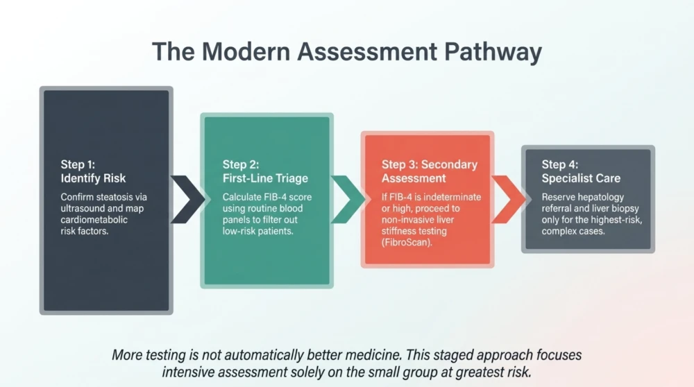
More testing is not automatically better medicine.
Tests have costs, limitations and false-positive or indeterminate results.
The objective is to use the right test in the right patient.
A staged approach helps concentrate additional testing on patients most likely to benefit from it.
Can Someone Have Advanced Fibrosis and Still Feel Completely Well?
Yes.
Chronic liver disease can remain clinically silent until relatively advanced stages.
This is precisely why relying on symptoms is problematic.
Absence of Symptoms Is Not a Fibrosis Test
A person does not need to have:
- Abdominal pain.
- Jaundice.
- Severe fatigue.
- Swelling.
- Loss of appetite.
to have clinically relevant fibrosis.
Symptoms become particularly important when advanced liver disease begins to decompensate, but our goal should be to identify high-risk disease before that point.
Can Bilirubin Remain Normal Too?
Yes.
Bilirubin can remain normal in compensated chronic liver disease.
Albumin and other markers of hepatic synthetic function may also remain preserved until disease becomes more advanced.
This is another reason the phrase “all my liver tests are normal” can be misleading when discussing fibrosis.
The Liver Has Considerable Functional Reserve
The liver can continue performing many of its essential functions despite chronic disease.
Therefore, routine biochemical tests may remain surprisingly reassuring while structural changes are developing.
This is one reason fibrosis assessment has become such an important part of modern MASLD management.
How Can Significant Fibrosis Develop When ALT Is Normal?
This question gets to the heart of why fatty liver with normal ALT can be misleading.
Many people expect liver disease to behave in a simple linear fashion:
More liver damage = higher ALT.
Less liver damage = lower ALT.
Unfortunately, chronic liver disease does not work that neatly.
ALT reflects leakage of an intracellular enzyme into the bloodstream during hepatocellular injury. Fibrosis, on the other hand, represents the accumulation and remodelling of extracellular scar tissue following repeated or persistent liver injury.
The two processes are related, but they are not proportional.
A patient can therefore have relatively little biochemical activity at the time of testing while carrying fibrosis accumulated over years.
Think of ALT as Activity and Fibrosis as Accumulated Damage
This is an imperfect analogy, but it can help patients understand the difference.
ALT provides information about current or recent hepatocellular injury.
Fibrosis provides information about scar tissue that has accumulated over time.
A normal ALT today therefore cannot tell us with certainty what has happened inside the liver over the previous 10 or 15 years.
Why Fibrosis Matters More Than the Ultrasound Grade
Patients often focus on whether an ultrasound says:
- Grade 1 fatty liver.
- Grade 2 fatty liver.
- Grade 3 fatty liver.
But this grading largely reflects the sonographic appearance of steatosis.
It is not the same as fibrosis staging.
Fat and Scar Tissue Are Different
Steatosis describes fat accumulation.
Fibrosis describes scar formation.
A conventional ultrasound cannot reliably distinguish all clinically important fibrosis stages.
This is why “mild fatty liver” on ultrasound should not automatically be translated into “mild liver disease.”
Can Severe Fatty Liver Exist Without Advanced Fibrosis?
Yes.
A person may have substantial steatosis but relatively little fibrosis.
The reverse issue is also important: the amount of visible steatosis does not provide a reliable direct measurement of the fibrosis burden.
Therefore, clinicians increasingly focus on identifying people at risk of advanced fibrosis rather than trying to predict prognosis from ultrasound steatosis grade alone.
Fibrosis Is a Major Prognostic Marker in MASLD
Across the MASLD spectrum, increasing fibrosis stage is strongly associated with worsening liver-related outcomes.
This makes fibrosis risk stratification one of the most clinically important parts of fatty-liver assessment.
The practical challenge is that most people with MASLD will not develop advanced liver disease.
We therefore need a way to separate the large lower-risk group from the smaller group requiring more intensive assessment.
This Is Why the Assessment Usually Happens in Steps
A practical pathway often looks like this:
Step 1: Identify steatotic liver disease or a person at increased metabolic risk.
Step 2: Assess cardiometabolic risk factors and alternative causes/contributors.
Step 3: Calculate a first-line fibrosis risk score such as FIB-4.
Step 4: If FIB-4 does not clearly indicate low risk, perform an appropriate second-line non-invasive fibrosis assessment.
Step 5: Refer or investigate further when results suggest advanced fibrosis, are discordant or remain clinically concerning.
This staged approach avoids sending every patient with fatty liver for invasive testing while reducing the chance of missing clinically important fibrosis.
Understanding FIB-4 Properly
FIB-4 uses four readily available variables:
- Age.
- AST.
- ALT.
- Platelet count.
It is inexpensive because these measurements are often already available from routine blood testing.
FIB-4 Is Particularly Useful for Ruling Out Advanced Fibrosis in Lower-Risk Results
Its major strength in primary-care pathways is helping identify people with a low probability of advanced fibrosis who may not need immediate specialist investigation.
Its purpose is not to tell us the exact histological fibrosis stage.
What Does a FIB-4 Below 1.3 Mean?
In many adults within the usual age range in which the score performs reasonably well, a FIB-4 below 1.3 is commonly used as a lower-risk threshold for advanced fibrosis.
It does not mean: “There is definitely zero fibrosis.”
It means the probability of advanced fibrosis is sufficiently low in the appropriate clinical context that immediate secondary testing may not be required.
What Does FIB-4 of 1.3 or Higher Mean?
It does not mean advanced fibrosis has been diagnosed.
It means FIB-4 has not confidently placed the patient in the low-risk category.
A second-line assessment is usually considered.
Common options include:
- Vibration-controlled transient elastography.
- ELF blood testing where available.
- Other validated elastography or fibrosis-assessment methods depending on the clinical setting.
Why FIB-4 Can Be Misleading in Younger Adults
Age is built directly into the FIB-4 calculation.
In adults younger than approximately 35 years, FIB-4 has lower accuracy for detecting advanced fibrosis.
A young patient with substantial metabolic risk should therefore not necessarily be dismissed solely because FIB-4 is low.
Consider the Whole Clinical Picture
A 31-year-old with severe obesity, type 2 diabetes, high triglycerides and substantial hepatic steatosis may warrant further assessment despite a reassuring-looking FIB-4.
Why FIB-4 Can Run Higher in Older Adults
The opposite problem occurs with ageing.
Because age contributes to the calculation, FIB-4 tends to increase as people get older.
For adults over approximately 65 years, a higher age-adjusted lower-risk threshold is commonly used to reduce unnecessary false-positive assessments.
Do Not Calculate FIB-4 During Acute Illness and Treat It as Chronic Fibrosis Staging
AST and ALT can change substantially during acute illness or acute hepatic injury.
A score calculated under those circumstances may not represent the person's stable chronic MASLD risk.
The clinical setting always matters.
Can Platelet Count Provide a Clue?
Yes.
Platelet count is part of FIB-4 for a reason.
As chronic liver disease progresses, particularly when portal hypertension develops, platelet count may decline.
However, thrombocytopenia has many other possible causes.
A low platelet count should therefore be investigated rather than automatically attributed to liver fibrosis.
What Is FibroScan?
FibroScan is a widely recognised device that uses vibration-controlled transient elastography to assess liver stiffness non-invasively.
The test is rapid and does not require needles or a liver biopsy.
Two measurements often attract particular attention:
- Liver stiffness measurement (LSM).
- Controlled attenuation parameter (CAP).
What Does Liver Stiffness Measurement Tell Us?
LSM is usually reported in kilopascals, or kPa.
Increasing stiffness can indicate increasing probability of fibrosis.
But it is essential to understand that liver stiffness is not identical to fibrosis.
FibroScan Does Not Literally Count Scar Tissue
It measures a physical property of the liver.
Fibrosis is a major determinant of liver stiffness, but other factors can influence the measurement.
Therefore, a particular kPa value should be interpreted within the clinical context and according to validated disease-specific thresholds.
What Can Temporarily Increase Liver Stiffness?
Potential confounders can include:
- Active hepatic inflammation.
- Cholestasis.
- Hepatic congestion.
- Recent food intake.
- Certain acute liver conditions.
Technical factors and severe obesity can also affect test reliability.
This is why proper preparation, probe selection and quality criteria matter.
What FibroScan Numbers Are Used in MASLD?
Exact interpretation varies according to the pathway and patient population.
However, contemporary MASLD pathways often use lower liver-stiffness values to help rule out advanced fibrosis and higher values to increase concern for advanced fibrosis.
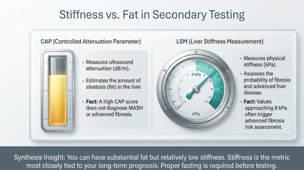
A commonly encountered threshold around 8 kPa is used in several pathways as a useful lower cut-off for advanced fibrosis risk assessment after an indeterminate or elevated FIB-4.
Values substantially above this require progressively greater attention.
Do Not Turn One kPa Value Into an Exact Fibrosis Stage
A FibroScan result should not be interpreted as:
“7.9 means no fibrosis and 8.1 means definite advanced fibrosis.”
Biological measurements do not work with that degree of precision.
Thresholds are decision points used to guide risk stratification.
What Is CAP?
CAP stands for controlled attenuation parameter.
It estimates ultrasound attenuation within the liver and is used as a non-invasive marker of hepatic steatosis.
CAP and liver stiffness answer different questions.
| FibroScan Measurement | Main Purpose |
|---|---|
| CAP | Estimates hepatic steatosis |
| Liver stiffness (kPa) | Assesses probability of fibrosis/advanced liver disease |
Can CAP Tell Me Whether I Have MASH?
No.
CAP provides information about steatosis.
It does not directly diagnose the histological inflammatory and hepatocellular injury features required to define MASH.
This distinction matters because patients sometimes assume that a high CAP score means severe MASH.
That conclusion cannot be made from CAP alone.
Can CAP Tell Me Whether My Liver Is Scarred?
No.
That is why the stiffness measurement is considered separately.
A patient can have substantial steatosis but relatively low stiffness.
Another patient may have less dramatic steatosis but clinically important fibrosis.
Why Fasting Before FibroScan Matters
Food intake can transiently affect liver-stiffness measurements.
Patients are therefore generally instructed to fast for an appropriate period before transient elastography according to the centre's protocol.
Following preparation instructions improves the interpretability of the examination.
What Is the ELF Test?
ELF stands for Enhanced Liver Fibrosis.
It is a blood-based test that combines biomarkers related to extracellular matrix turnover.
It can be used as a second-line non-invasive fibrosis assessment in appropriate pathways.
ELF Is Different From ALT and AST
ALT and AST primarily reflect hepatocellular injury.
ELF is designed around biomarkers related to fibrosis biology.
It therefore addresses a different clinical question.
What About MR Elastography?
Magnetic resonance elastography, or MRE, uses MRI technology to assess tissue stiffness.
It has high diagnostic performance for liver fibrosis.
However, cost, availability and the need for specialised equipment mean it is generally not the first test performed for every patient with fatty liver.
What Is MRI-PDFF?
MRI-proton density fat fraction can quantify liver fat with high accuracy.
It is particularly useful in research and selected specialist situations.
But quantifying liver fat precisely is not the same as staging fibrosis.
More Liver Fat Does Not Automatically Mean More Fibrosis
These processes are related but distinct.
This is another reason fatty-liver management should not revolve around a single number.
Does Anyone Still Need a Liver Biopsy?
Yes, but far fewer patients need biopsy than in the past.
Modern non-invasive tests can stratify fibrosis risk effectively in many people.
Liver biopsy may still be considered when:
- Non-invasive tests are indeterminate.
- Different tests provide conflicting results.
- There is concern for significant disease that cannot otherwise be clarified.
- An alternative or additional liver disorder is suspected.
- Histological confirmation would materially change management.
Why Is Liver Biopsy Not Used for Everyone?
Biopsy is invasive.
It carries procedural risks.
It samples only a tiny portion of the liver.
And when non-invasive testing can answer the clinical question sufficiently well, exposing every person with fatty liver to biopsy would be unnecessary.
Type 2 Diabetes Deserves Special Attention
If there is one metabolic condition that should make clinicians particularly alert to fibrosis risk in MASLD, it is type 2 diabetes.
People with type 2 diabetes have a substantially higher prevalence of MASLD and are at greater risk of progressive liver disease.
Normal ALT Should Not Cancel Fibrosis Assessment in a High-Risk Patient
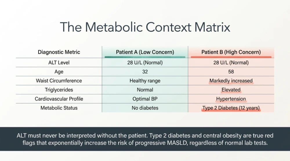
Consider two patients.
| Feature | Patient A | Patient B |
|---|---|---|
| ALT | Normal | Normal |
| Age | 32 | 58 |
| Type 2 diabetes | No | Yes, 12 years |
| Waist circumference | Healthy range | Markedly increased |
| Triglycerides | Normal | Elevated |
| Hypertension | No | Yes |
Both patients have normal ALT.
But their probability of clinically important metabolic liver disease is not the same.
This is why ALT should never be interpreted without the patient.
Why Does Diabetes Increase Liver Risk?
Type 2 diabetes and MASLD share important underlying mechanisms, including insulin resistance, altered adipose-tissue biology and abnormalities in lipid metabolism.
Diabetes is also associated with a greater probability of MASH and advanced fibrosis.
This relationship works in both directions: MASLD is also associated with increased future risk of developing type 2 diabetes.
Should Every Person With Diabetes Be Checked for Fibrosis?
Current major guidance increasingly supports fibrosis risk assessment in people with type 2 diabetes, particularly when MASLD or other metabolic risk factors are present.
The purpose is not to send every patient directly to hepatology.
It is to identify those who need further evaluation while managing lower-risk patients appropriately in primary or metabolic care.
What About Prediabetes?
Prediabetes is another marker of metabolic dysfunction.
When prediabetes occurs alongside central obesity, high triglycerides, hypertension and fatty liver, the combined metabolic phenotype deserves attention.
Again, ALT may remain normal throughout this period.
Can Significant Fibrosis Occur in Someone Without Diabetes?
Yes.
Diabetes increases risk, but it is not required for fibrosis progression.
Other factors can contribute, including:
- Increasing age.
- Obesity and visceral adiposity.
- Multiple cardiometabolic risk factors.
- Genetic susceptibility.
- Alcohol exposure.
- Coexisting liver disease.
Alcohol Still Matters in Someone With Metabolic Fatty Liver
The newer steatotic liver disease terminology recognises that metabolic dysfunction and alcohol exposure can coexist.
It is therefore important to obtain an accurate alcohol history rather than assuming that every fatty liver in someone with obesity is exclusively metabolic.
Metabolic Risk Does Not Make Alcohol Irrelevant
Alcohol and metabolic risk factors can interact adversely.
Patients should provide an accurate estimate of the type, amount and frequency of alcohol consumed so that liver disease can be classified and managed appropriately.
Do Not Forget Other Causes of Liver Disease
Finding steatosis does not mean every abnormality should automatically be blamed on MASLD.
Depending on the patient, clinicians may need to consider other conditions such as:
- Viral hepatitis.
- Alcohol-related liver disease.
- Medication-related liver injury.
- Autoimmune liver disease.
- Iron overload disorders.
- Other metabolic or genetic liver diseases.
The extent of testing should be guided by history, examination, age, biochemical abnormalities and clinical probability.
Can Weight Loss Improve Fatty Liver?
Yes.
When excess adiposity is present, sustained weight reduction can improve hepatic steatosis and several associated metabolic abnormalities.
Greater sustained weight loss is generally associated with greater probability of improving more advanced features of metabolic liver disease.
But Do Not Wait for Major Weight Loss Before Exercising
Physical activity can improve cardiometabolic health and may reduce liver fat even when weight loss is modest.
This is an important message for patients who begin exercising but become discouraged because the weighing scale changes slowly.
Resistance Training or Cardio for Fatty Liver?
Both can be useful.
Aerobic activity improves cardiorespiratory fitness and can reduce liver fat.
Resistance training helps preserve and improve skeletal muscle, which is important for glucose disposal and metabolic health.
For many patients, combining both is preferable to searching for one perfect form of exercise.
Muscle Matters in Fatty Liver Disease
Skeletal muscle is metabolically active tissue.
Low muscle mass and poor muscle function can coexist with obesity, particularly as people age.
This combination can create an unfavourable metabolic phenotype even when total body weight appears unchanged.
Do Not Treat MASLD With Weight Loss Alone
The objective should usually be to reduce excess fat while preserving muscle and improving physical fitness.
A rapidly falling scale accompanied by substantial muscle loss is not an ideal metabolic outcome.
What Should Be Monitored Besides ALT?
| Assessment | What It Adds |
|---|---|
| Weight and waist circumference | Tracks excess and central adiposity |
| Blood pressure | Assesses associated cardiovascular risk |
| Fasting glucose/HbA1c | Assesses dysglycaemia and diabetes risk |
| Lipid profile | Identifies associated dyslipidaemia and cardiovascular risk |
| AST, ALT and platelet count | Contribute to liver assessment and FIB-4 calculation |
| FIB-4 | First-line advanced-fibrosis risk stratification |
| Transient elastography when indicated | Provides secondary assessment of liver stiffness/fibrosis risk |
| Cardiovascular risk assessment | Addresses a major source of morbidity in MASLD |
Why Cardiovascular Risk Cannot Be Ignored
Patients understandably focus on whether fatty liver will eventually cause cirrhosis.
That risk matters, particularly in people with advanced fibrosis.
But MASLD occurs within a broader cardiometabolic environment.
Blood pressure, diabetes, dyslipidaemia, smoking, physical inactivity and obesity all require appropriate management.
Treat the Patient, Not Just the Ultrasound
A successful MASLD management plan should not consist merely of repeating ALT and performing another ultrasound every year.
It should address the underlying metabolic and cardiovascular risk factors that frequently accompany the liver disease.
What if ALT Becomes Normal After Weight Loss?
That can be an encouraging sign.
Improving liver enzymes alongside weight reduction and better metabolic control may reflect improvement in hepatocellular injury.
But if a patient previously had evidence suggesting significant fibrosis, normalisation of ALT should not automatically terminate appropriate fibrosis follow-up.
Can Fibrosis Improve?
Yes.
Liver fibrosis is not necessarily a one-way process.
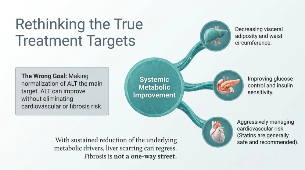
When the underlying metabolic and hepatic injury is substantially reduced, fibrosis can regress in some patients.
This is another reason early identification of higher-risk disease matters.
Can Cirrhosis Be Present With Normal ALT?
Yes.
Aminotransferase levels can be normal in people with advanced chronic liver disease, including cirrhosis.
This is one of the clearest demonstrations of why ALT should never be used as a stand-alone fibrosis test.
What Other Clues May Suggest Advanced Liver Disease?
Depending on the stage and cause, clinicians may encounter findings such as:
- Low platelet count.
- Splenomegaly.
- Low albumin.
- Elevated bilirubin.
- Prolonged INR.
- Ascites.
- Oesophageal or gastric varices.
- Hepatic encephalopathy.
However, waiting for these abnormalities to appear would mean waiting until disease is much more advanced.
The purpose of non-invasive fibrosis assessment is to identify risk earlier.
How Should Fatty Liver With Normal ALT Be Managed?
Once we understand that fatty liver with normal ALT does not automatically mean harmless liver disease, the next question becomes practical:
“What should I actually do?”
The answer depends on two related but different risks:
- The risk of progressive liver disease, particularly advanced fibrosis.
- The person's overall cardiometabolic risk, including obesity, diabetes, dyslipidaemia and hypertension.
For many patients, treatment is therefore not simply about “reducing liver fat.”
It is about improving the metabolic environment that contributed to fatty liver while identifying the smaller group who already have clinically important fibrosis.
Start by Establishing the Fibrosis Risk
If fatty liver has been identified, particularly in someone with type 2 diabetes, obesity or multiple cardiometabolic risk factors, simply repeating ALT every few months is not an adequate fibrosis strategy.
A structured assessment can include:
- Medical and alcohol history.
- Medication review.
- Weight and waist circumference.
- Blood pressure.
- Glucose status and HbA1c.
- Lipid profile.
- AST and ALT.
- Platelet count.
- FIB-4 calculation.
- Secondary non-invasive fibrosis testing when indicated.
- Assessment for alternative or additional liver disease when clinically appropriate.
The exact pathway should be individualised.
Do Not Treat the ALT Number—Treat the Patient
One of the biggest mistakes in fatty-liver management is making normalisation of ALT the main treatment target.
ALT can certainly be followed.
But a patient can improve ALT without eliminating all liver or cardiovascular risk.
Conversely, meaningful metabolic improvements may occur without dramatic changes in ALT if ALT was already normal.
Better Questions Include:
- Is excess body fat decreasing?
- Is waist circumference decreasing?
- Is glucose control improving?
- Are triglycerides improving?
- Is blood pressure controlled?
- Is physical fitness improving?
- Has fibrosis risk been assessed appropriately?
- Is alcohol exposure relevant?
Weight Reduction Can Produce Major Liver Benefits
When excess adiposity is present, sustained weight loss is one of the most effective interventions for MASLD.
As weight decreases, visceral and liver fat can decrease substantially.
Insulin sensitivity may improve.
Triglycerides and glucose regulation may improve.
And with sufficient sustained weight reduction, inflammatory liver disease and fibrosis can improve in some patients.
How Much Weight Loss Should You Aim For?
There is no single number appropriate for every patient.
However, a useful general principle is that liver benefits tend to increase as sustained weight loss becomes greater.
Even modest weight reduction can reduce hepatic steatosis.
Approximately 5% weight loss can provide meaningful improvement in liver fat in many patients.
Greater reductions—often in the range of 7–10% or more—are generally associated with a greater likelihood of improving steatohepatitis and other metabolic abnormalities.
In patients with obesity and more advanced metabolic liver disease, weight reduction beyond 10% may provide additional benefit when achieved safely and maintained.
These Are Treatment Targets, Not Pass-or-Fail Numbers
A patient who loses 6% of body weight has not “failed” because they did not reach 10%.
Similarly, someone who needs substantial obesity treatment should not stop at 5% simply because liver fat has started improving.
The appropriate target depends on obesity severity, diabetes, liver fibrosis, physical function and other medical conditions.
For an 80 kg Person, What Do These Percentages Mean?
| Weight Reduction | Approximate Weight Loss From 80 kg | Potential Significance |
|---|---|---|
| 5% | 4 kg | Can meaningfully reduce hepatic steatosis and improve metabolic health |
| 7% | 5.6 kg | Greater metabolic and liver benefit may occur |
| 10% | 8 kg | Associated with greater likelihood of improvement in MASH and fibrosis-related outcomes |
But Weight Loss Should Not Mean Muscle Loss
This deserves more attention than it usually receives.
If a patient loses 10 kg but a substantial proportion comes from skeletal muscle, the weighing scale may look successful while physical reserve deteriorates.
Muscle is metabolically important.
It is a major site of glucose disposal and is essential for strength, mobility and healthy ageing.
The Better Goal Is Fat Loss With Muscle Preservation
A well-designed programme generally combines:
- An appropriate energy deficit when weight reduction is needed.
- Adequate protein according to individual requirements.
- Resistance training.
- Aerobic activity.
- Regular everyday movement.
- Adequate recovery and sleep.
What Should You Eat for Fatty Liver?
There is no single “fatty liver diet.”
What matters most is a sustainable dietary pattern that improves overall diet quality and, when appropriate, creates sufficient energy deficit to reduce excess adiposity.
Mediterranean-style dietary patterns have particularly strong cardiometabolic rationale in MASLD.
But this does not mean an Indian patient must suddenly start eating an unfamiliar European menu.
A Mediterranean-Style Pattern Can Be Adapted to Indian Food
The principles matter more than the geography.
- Eat plenty of vegetables.
- Include whole fruit rather than fruit juice.
- Use pulses and legumes regularly.
- Choose appropriate portions of minimally processed whole grains.
- Include nuts and seeds in calorie-appropriate quantities.
- Use unsaturated fats appropriately.
- Include adequate protein.
- Limit sugar-sweetened beverages.
- Reduce excessive refined carbohydrates.
- Limit highly processed foods.
- Avoid routinely consuming more calories than your body requires.
What Can an Indian Fatty-Liver Plate Look Like?
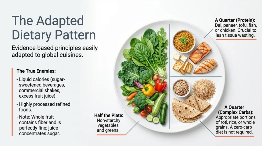
A practical meal might contain:
- A generous portion of non-starchy vegetables.
- Dal, rajma, chana, curd, paneer, tofu, eggs, fish, chicken or another suitable protein source.
- An appropriate portion of roti, rice or another staple carbohydrate.
- Salad or cooked vegetables.
- Whole fruit when desired rather than juice.
The precise proportions depend on energy requirements, glucose status, activity, dietary preference and weight-management goals.
Do You Need to Stop Eating Rice and Roti?
No.
Fatty liver is not treated simply by banning individual staple foods.
Total energy intake, portion size, carbohydrate quality, protein, fibre and the overall dietary pattern are more important.
A patient can overconsume calories on a low-carbohydrate diet just as easily as on a high-carbohydrate diet.
Reduce Added Sugar and Sugar-Sweetened Beverages
Sugar-sweetened beverages are particularly easy to overconsume because liquid calories provide relatively little satiety.
This includes:
- Soft drinks.
- Sweetened packaged beverages.
- Energy drinks.
- Sweetened tea or coffee consumed frequently.
- Many commercial shakes.
- Excessive fruit juice.
Whole Fruit Is Not the Same as Fruit Juice
Whole fruit contains fibre and requires chewing.
Fruit juice concentrates sugar and calories into a form that can be consumed rapidly.
For most people with MASLD, appropriate portions of whole fruit can remain part of a healthy diet.
Do You Need a Zero-Sugar Diet?
No.
The aim is not to become afraid of every gram of naturally occurring sugar.
The more useful goal is to reduce excessive added sugar and highly refined foods while improving overall diet quality and energy balance.
Protein Deserves Attention
Protein becomes particularly important when a patient with MASLD is deliberately losing weight.
An inadequate protein intake combined with aggressive calorie restriction can increase loss of lean tissue.
Protein requirements should be individualised according to body size, age, physical activity, calorie intake, kidney function and other medical conditions.
Common Indian Protein Options Include:
- Dal and pulses.
- Soya and tofu.
- Milk and curd where appropriate.
- Paneer in suitable portions.
- Eggs.
- Fish.
- Chicken and other lean meats.
- Protein supplementation when genuinely needed and clinically appropriate.
Exercise Can Help Even Before Major Weight Loss Occurs
This is one of the most encouraging aspects of MASLD management.
Regular physical activity can improve insulin sensitivity, cardiorespiratory fitness and liver fat even when the weighing scale changes relatively little.
Exercise therefore has value beyond simply burning calories.
How Much Exercise?
For most adults, a practical target is to progressively work towards approximately 150–300 minutes of moderate-intensity aerobic activity per week, or an appropriate equivalent of vigorous activity.
Muscle-strengthening exercise should also be included on at least two days per week where medically appropriate.
Start Below the Target if Necessary
A sedentary person does not need to reach 300 minutes in the first week.
Beginning with regular walking and gradually increasing duration and intensity can be much more sustainable.
Walking After Meals Is Particularly Practical
A short walk after meals can improve post-meal glucose handling and increase daily activity.
For patients with insulin resistance, prediabetes or type 2 diabetes, this can be a useful habit alongside the broader exercise programme.
Resistance Training Should Not Be Forgotten
Fatty-liver management is sometimes reduced to “walk more and lose weight.”
That misses the importance of skeletal muscle.
Resistance training can improve strength, preserve lean mass during weight loss and support glucose metabolism.
For many adults, combining aerobic exercise with resistance training is preferable to relying exclusively on cardio.
What About Alcohol?
Alcohol intake deserves an individual discussion in anyone with steatotic liver disease.
The idea that a particular amount of alcohol is automatically “safe for the liver” regardless of fibrosis stage and metabolic risk is too simplistic.
Alcohol exposure and metabolic liver injury can coexist.
Advanced Fibrosis Changes the Conversation
Patients with significant fibrosis or cirrhosis generally require much stricter alcohol advice, and abstinence may be recommended.
For other patients, alcohol advice should take into account the amount consumed, pattern of drinking, fibrosis risk and overall medical context.
What About Coffee?
Observational evidence has repeatedly associated coffee consumption with lower risk of several chronic liver outcomes.
For adults who already tolerate coffee and have no reason to avoid caffeine, moderate unsweetened coffee may fit within an overall healthy dietary pattern.
But coffee should not be sold as a treatment for fibrosis.
Do Not Turn Coffee Into a High-Calorie Dessert
A coffee containing large quantities of sugar, cream or flavoured syrup is metabolically very different from plain or lightly prepared coffee.
Do Liver Detox Drinks Help?
There is no need to “flush” fat or toxins out of the liver using juices, herbal cleanses or detox products.
The liver is itself one of the body's major metabolic and detoxification organs.
What it needs is reduction of the underlying drivers of injury—not a commercial cleanse.
Be Careful With Herbal and “Natural” Liver Supplements
Natural does not automatically mean safe.
Some herbal, bodybuilding and weight-loss supplements can cause drug-induced liver injury.
Patients should tell their doctor about supplements they are taking rather than assuming that a product labelled “liver support” must protect the liver.
What About Vitamin E?
Vitamin E has been studied in selected patients with steatohepatitis, but it is not a universal supplement for everyone with ultrasound-detected fatty liver.
Potential benefits and risks depend on the patient population and clinical indication.
It should therefore not be started routinely simply because ALT or ultrasound is abnormal.
Diabetes Treatment Is Part of Liver Treatment
When type 2 diabetes and MASLD coexist, glucose management is not a separate issue.
These diseases share important metabolic drivers.
Some glucose-lowering medications can also improve body weight and certain liver-related outcomes, making treatment selection particularly relevant in the appropriate patient.
What About GLP-1-Based Treatment?
GLP-1 receptor agonist and related incretin-based therapies can produce substantial weight loss in appropriately selected patients with obesity and can improve several cardiometabolic abnormalities.
Some agents also have evidence for improvement in steatotic liver disease outcomes.
However, the decision to prescribe them should be based on approved indications, the individual's obesity and diabetes status, contraindications, adverse-effect profile and broader clinical context.
There Are Now Disease-Directed Treatments for Selected MASH Patients
The treatment landscape for MASH has changed significantly.
In selected patients with non-cirrhotic MASH and clinically important fibrosis, disease-directed pharmacological treatment may now be considered depending on regulatory approval, availability and specialist assessment.
This makes accurate fibrosis assessment even more relevant than it was when lifestyle modification was essentially the only disease-specific strategy.
Not Every Person With Fatty Liver Needs a MASH Drug
An incidental ultrasound showing steatosis with low fibrosis risk is very different from non-cirrhotic MASH with moderate-to-advanced fibrosis.
Treatment should follow disease severity rather than the presence of liver fat alone.
Statins and Fatty Liver: A Common Misunderstanding
Patients with fatty liver frequently have high LDL cholesterol or other cardiovascular risk factors.
Some avoid statins because they believe any liver condition makes cholesterol-lowering therapy unsafe.
This is generally incorrect.
Statins can be used in most people with MASLD when clinically indicated for cardiovascular risk reduction.
Do Not Leave Cardiovascular Risk Untreated Because of Fatty Liver
For many people with MASLD, preventing cardiovascular disease is a major component of long-term care.
The presence of fatty liver should prompt appropriate cardiovascular risk management rather than distract from it.
How Often Should FIB-4 Be Repeated?
The interval depends on metabolic risk.
Patients with higher-risk metabolic profiles, particularly type 2 diabetes or multiple cardiometabolic risk factors, generally require reassessment more frequently than someone with fewer risk factors and a clearly low-risk initial assessment.
Follow-up intervals should therefore be individualised rather than applying one annual test to everyone.
Does FibroScan Need to Be Repeated Every Year?
Not automatically.
The need for repeat transient elastography depends on:
- The initial liver-stiffness result.
- FIB-4 trend.
- Diabetes and other metabolic risk factors.
- Changes in weight and metabolic health.
- Whether previous results were borderline or discordant.
- Whether the result would alter management.
More frequent testing does not necessarily produce better care.
A Practical Monitoring Framework
| What to Monitor | Why It Matters |
|---|---|
| Weight trend | Tracks progress when excess adiposity is being treated |
| Waist circumference | Provides practical information about central adiposity |
| Blood pressure | Important cardiovascular and metabolic risk factor |
| Glucose/HbA1c | Identifies and monitors dysglycaemia |
| Lipid profile | Assesses dyslipidaemia and cardiovascular risk |
| AST and ALT | Provides biochemical information but does not independently stage fibrosis |
| Platelet count | Contributes to FIB-4 and may provide additional context in advanced liver disease |
| FIB-4 | Helps stratify advanced-fibrosis risk |
| Transient elastography when indicated | Provides secondary assessment of liver stiffness |
| Strength and fitness | Tracks important metabolic and functional improvements |
When Should You See a Liver Specialist?
Specialist assessment may be appropriate when non-invasive tests suggest increased risk of advanced fibrosis, results are repeatedly indeterminate or conflicting, another liver disease is suspected, or there are features of advanced chronic liver disease.
Referral decisions should consider the entire clinical picture rather than ALT alone.
Red Flags That Need Prompt Medical Assessment
Seek medical assessment promptly if chronic liver disease is accompanied by features such as:
- New jaundice.
- Increasing abdominal swelling.
- Vomiting blood.
- Black, tarry stools.
- New confusion or unusual drowsiness.
- Marked swelling of the legs.
- Unexplained significant weight loss.
- Persistent severe abdominal symptoms.
These symptoms are not typical markers of uncomplicated early fatty liver and can indicate more serious disease requiring timely evaluation.
Common Myths About Fatty Liver With Normal ALT
Myth 1: Normal ALT Means My Liver Is Healthy
False.
Normal ALT is useful information but cannot exclude MASLD, MASH or clinically significant fibrosis.
Myth 2: Grade 1 Fatty Liver Means Stage 1 Fibrosis
False.
Ultrasound steatosis grade and fibrosis stage are different concepts.
Myth 3: If AST and ALT Are Normal, I Do Not Need Fibrosis Assessment
Not necessarily.
Fibrosis assessment depends on overall risk, particularly diabetes, obesity and other cardiometabolic factors.
Myth 4: FibroScan and Ultrasound Are the Same Test
No.
Conventional ultrasound mainly identifies structural abnormalities and can suggest steatosis. Transient elastography provides information about liver stiffness and therefore fibrosis risk.
Myth 5: A High CAP Score Means Advanced Fibrosis
False.
CAP primarily estimates steatosis. Liver stiffness is the FibroScan measurement more directly relevant to fibrosis risk.
Myth 6: FIB-4 Above 1.3 Means I Have Advanced Fibrosis
False.
In common adult pathways, this is a threshold for further assessment rather than a diagnosis of advanced fibrosis.
Myth 7: Only People With Obesity Develop Fatty Liver
False.
MASLD can occur at lower BMI, including in South Asian populations.
Myth 8: Liver Detox Drinks Can Remove Fat From the Liver
There is no convincing evidence that commercial detox drinks remove hepatic fat or reverse fibrosis.
Myth 9: Fatty Liver Means I Must Stop Eating All Carbohydrates
False.
The overall dietary pattern, calorie balance, carbohydrate quality, fibre and protein intake matter more than eliminating an entire macronutrient.
Myth 10: Nothing Can Be Done Once Fibrosis Starts
False.
Fibrosis can regress in some patients when the underlying liver injury and metabolic drivers are substantially improved. The likelihood depends on disease stage and treatment response.
What I Tell Patients With Fatty Liver and Normal ALT
If you show me an ultrasound reporting fatty liver and your ALT is normal, I consider that ALT result reassuring in one limited sense: there is no biochemical ALT elevation at that moment.
But I would not use it to determine whether your liver contains significant scar tissue.
I would look at your age, weight, waist circumference, glucose status, diabetes history, triglycerides, blood pressure, AST, ALT and platelet count.
I would calculate or review your fibrosis risk where appropriate.
If the first-line assessment is reassuring, you may not need extensive liver investigations.
If it is not clearly reassuring, the next step may be an appropriate non-invasive fibrosis test such as transient elastography.
This approach avoids both extremes:
Ignoring fatty liver because ALT is normal. And: Sending every person with fatty liver for extensive investigations.
A Practical Fatty-Liver Checklist
If your scan reports fatty liver, ask:
- What is my waist circumference?
- Do I have prediabetes or type 2 diabetes?
- What are my triglycerides and HDL cholesterol?
- Is my blood pressure controlled?
- What are my AST and ALT?
- What is my platelet count?
- Has my FIB-4 been assessed where appropriate?
- Do I need secondary fibrosis assessment?
- Is my alcohol intake relevant?
- Could another liver disease be present?
- Do I need weight reduction?
- Am I preserving muscle while losing weight?
- Am I getting regular aerobic activity?
- Am I doing resistance training?
- Are my diabetes and cardiovascular risks being treated appropriately?
Key Takeaways
- Fatty liver with normal ALT is common and should not automatically be considered harmless.
- ALT is primarily a biochemical marker of hepatocellular injury; it does not directly measure liver fibrosis.
- Normal ALT and AST cannot reliably exclude clinically significant fibrosis.
- The ultrasound grade of steatosis is not the same as the fibrosis stage.
- Fibrosis burden is one of the most important determinants of long-term liver-related risk in MASLD.
- FIB-4 is commonly used as a first-line non-invasive assessment for advanced-fibrosis risk.
- A FIB-4 threshold such as 1.3 is a decision point for further assessment in appropriate adults—not a diagnosis of advanced fibrosis.
- Age affects FIB-4 interpretation.
- Type 2 diabetes substantially increases concern for progressive MASLD and fibrosis.
- Transient elastography can provide additional information about liver stiffness when secondary assessment is indicated.
- CAP estimates steatosis and should not be confused with fibrosis measurement.
- Not everyone with fatty liver requires FibroScan, MRI or liver biopsy.
- When excess adiposity is present, sustained weight reduction can substantially improve liver and metabolic health.
- Exercise can reduce liver fat and improve metabolic health even before major weight loss occurs.
- Resistance training is valuable because preserving muscle is part of good metabolic treatment.
- MASLD management should also address diabetes, blood pressure, lipids, sleep, alcohol exposure and cardiovascular risk.
- A normal ALT should never replace appropriate fibrosis risk assessment in a high-risk patient.
References
- European Association for the Study of the Liver, European Association for the Study of Diabetes and European Association for the Study of Obesity. EASL-EASD-EASO Clinical Practice Guidelines on the Management of Metabolic Dysfunction-Associated Steatotic Liver Disease. Journal of Hepatology. 2024.
- American Association for the Study of Liver Diseases. Clinical guidance and educational resources concerning MASLD, fibrosis risk stratification and non-invasive liver disease assessment.
- Rinella ME, et al. AASLD Practice Guidance on the Clinical Assessment and Management of Nonalcoholic Fatty Liver Disease. Hepatology. 2023. Guidance remains an important evidence base for non-invasive fibrosis assessment, metabolic risk evaluation and management.
- European Association for the Study of the Liver. Clinical guidance concerning non-invasive tests for evaluation of liver disease severity and prognosis.
- World Health Organization. WHO Guidelines on Physical Activity and Sedentary Behaviour. Recommendations concerning aerobic activity, muscle-strengthening exercise and sedentary behaviour.
- Peer-reviewed longitudinal studies evaluating fibrosis stage and long-term clinical outcomes in steatotic liver disease.
- Peer-reviewed systematic reviews and meta-analyses evaluating weight reduction, aerobic exercise, resistance training and dietary interventions in MASLD.
- Peer-reviewed studies evaluating FIB-4, vibration-controlled transient elastography, ELF and magnetic resonance elastography for non-invasive assessment of liver fibrosis.
Medical note: This article provides general health information and does not replace individual medical assessment. A normal ALT cannot independently determine the severity of fatty liver disease. Fibrosis assessment, additional investigations and follow-up should be selected according to age, metabolic risk, medical history, medications, alcohol exposure and the results of validated non-invasive tests.
Written by Dr. Pankaj Kumar , General & Lifestyle Physician, Dwarka, New Delhi